What is Filopodia? What are Filopodia?
What are they?
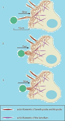
Filopodia are thin, dynamic cell extensions comprising tight bundles of long actin filaments covered with cell membrane. They can change their length rapidly, span many cell diameters, and interact with other cells. Sometimes the name filopodia is used more broadly to describe a range of cytoskeletal protrusions including thick filopodia, cell feet, and amoebae pseudopods.
These structures can be more stable or thicker than canonical filopodia, and may also contain microtubules. They first came to prominence… In the 1960s when they were shown to be involved in sea urchin gastrulation. During the invagination of the sea urchin endoderm, mesenchymal cells extend filopodia to ectodermal cells across the blastocoel cavity; these filopodia are responsible for the directed migration of the mesenchyme.
Which cells have them?
Which cells have them?
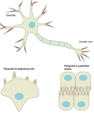
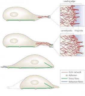
Many types of motile cell use filopodia. They are often associated with lamellipodia, sheet-like webs of actin filaments, at the advancing edge of migrating cells such as fibroblasts and keratinocytes. In neurons, they are clearly seen in growth cones, and dendritic spines are essentially short filopodia. Sometimes they are difficult to see because their delicate structure can be damaged by fixation but, as live microscopy improves, the number of tissues where they can be detected is increasing.
What is Filopodia? What are Filopodia?
What do they do?
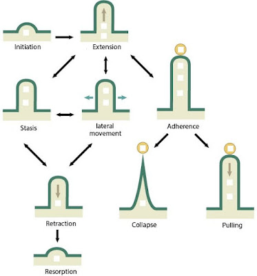
Filopodia seem to explore the extracellular matrix and surfaces of other cells, identify appropriate targets for adhesion, and then generate guidance cues and traction forces to move the cell body. These functions are required in cell migration and many morphogenetic events, including gastrulation, axonal pathfinding, epithelial cell adhesion, dorsal closure in Drosophila, ventral enclosure in Caenorhabditis elegans, and wound healing.
How thick, how long, how fast, how strong?
The classical sea urchin filopodia are long, thin and straight, 0.2 to 0.4 µm in diameter, and normally extend 5 to 35 µm, though occasionally they can be more than 70 µm in length. Normally they grow at rates up to 10 µm/min, with bursts of maximum activity up to 25 µm/min. Their retraction rate is similar and they tend to kink during that process as if the disassembly destabilizes the structure. Filopodia are quite flexible and capable of waving around searching for the right targets but are also very strong as their structural integrity is maintained even when they extend over 70 µm.
What about cytonemes?
What about cytonemes?
Some types of filopodia have exclusive names. Cytonemes are up to 800 µm long and are found in Drosophila wing imaginal disc. They may be involved in long-range cell-cell communication. Myopodia protrude from differentiating muscle cells and intermingle with growth cone filopodia, clustering at the site of motor neuron innervation.
How do they form?
How do they form?
Filopodia extend via actin polymerization at their tips. It is not clear if there is a universal mechanism for filopodia induction but in several cell lines, the process seems to be triggered by the binding of the small GTPase Cdc42 to N-WASP, an essential regulator of actin cytoskeleton formation. Activated N-WASP binds to Profilin and Arp2/3 and this complex nucleates the formation of a new filopodium.
What controls their behavior?
What controls their behavior?
The interaction of filopodia with their targets can elicit attraction, repulsion, adhesion or disassembly. Some of the secreted and membrane-associated proteins that mediate these processes are known, however, the pathways that signal to the cytoskeleton machinery are largely unknown and their discovery is a challenge for the future.
To Know more....
What is Filopodia? What are Filopodia?
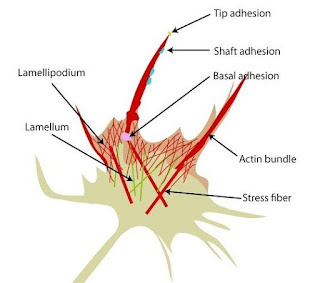
Filopodia additionally referred to as microspikes, square measure slender cytoplasmatic projections that reach on the far side the vanguard of lamellipodia in migrating cells.They contain actin filaments cross-linked into bundles by actin-binding proteins, e.g. fascin and fimbrin. Filopodia from focal adhesions with the substratum, linking it to the cell surface.
Activation of the Rho family of small Ras-related GTPases, particularly cdc42 and their downstream intermediates results in the polymerization of actin fibers by Ena/Vasp homology proteins.
Growth factors bind to receptor amino acid kinases leading to the polymerization of simple protein filaments, which, when cross-linked, make up the supporting cytoskeletal elements of filopodia.
Rho activity also results from inactivation by phosphorylation of ezrin-moesin-radixin family proteins that link actin filaments to the filopodia membrane.
To close a wound in vertebrates, growth factors stimulate the formation of filopodia in fibroblasts to direct fibroblast migration and wound closure.
In neurons deprived of filopodia by partial inhibition of actin filaments polymerization, growth cone extension continues as normal but the direction of growth is disrupted and highly irregular.
Filopodia-like projections have additionally been connected to nerve fiber creation once new synapses square measure fashioned within the brain.
Filopodia are also used for movement of bacteria between cells, so as to evade the host immune system. The intracellular bacteria Ehrlichia are transported between cells through the host cell filopodia induced by the pathogen during the initial stages of infection.
Viruses were shown to be transported on filopodia toward the cell body, leading to cell infection.
Directed transport of receptor-bound epidermic protein (EGF) on filopodia has additionally been delineate, supporting the planned sensing perform of filopodia.

0 Comments
please do not post spam link.